Ocular Imaging
Imaging the eye and retina to understand biological processes and disease progression
Research Focus
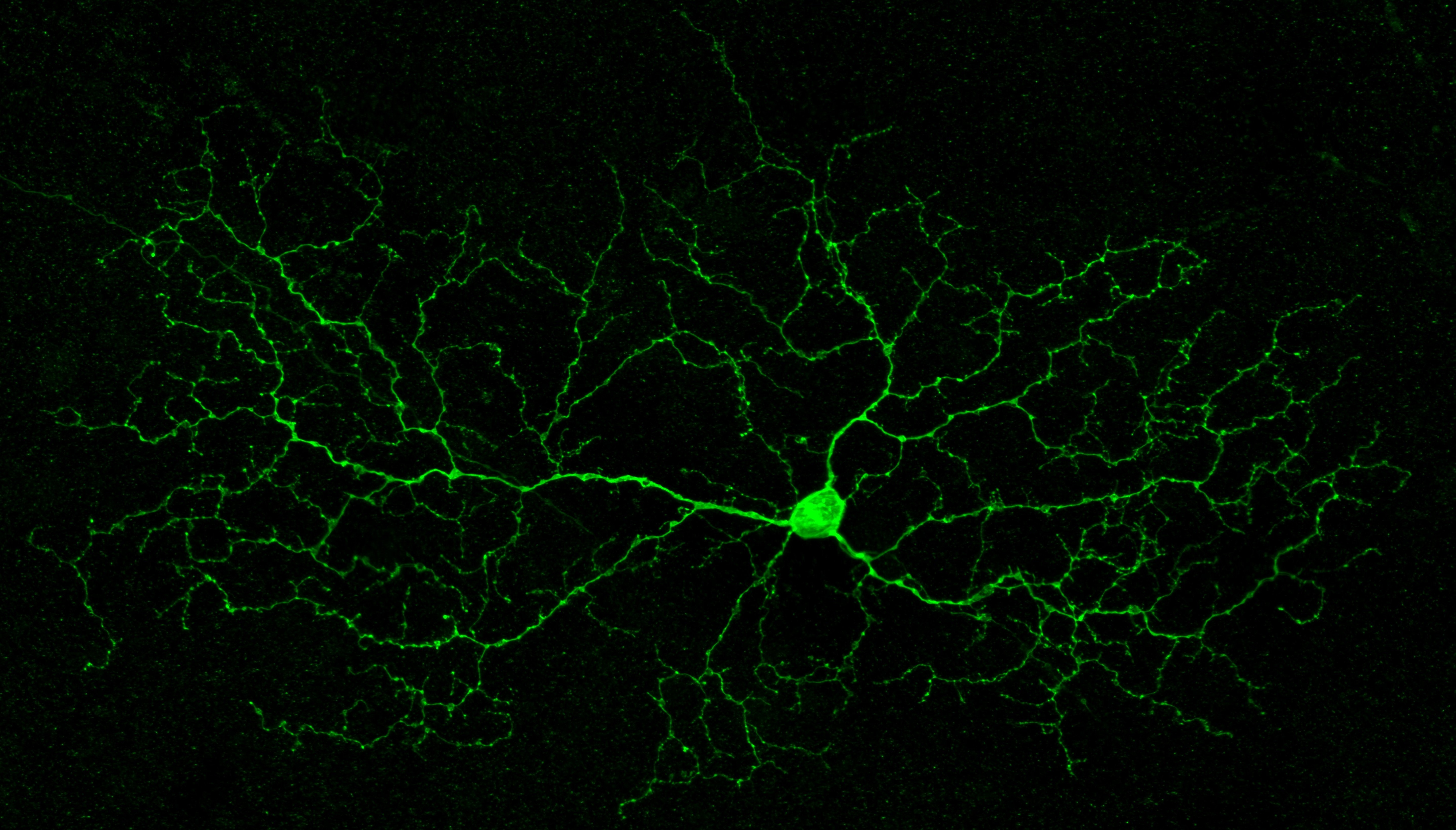
Imaging is central to the diagnosis of many eye diseases as well as understanding biological processes. These labs use diverse imaging techniques to understand eye disease and eye biology with ever greater resolution and precision.
Key Research Areas:
- Fundus imaging
- Optical coherence tomography
- Advanced microscopy
Faculty
Adrian Au
Assistant Professor
Imaging, AI, and Retinal Genomics
Dr. Au’s research leverages advanced ophthalmic imaging, retinal genomics, and artificial intelligence to improve disease prognostication and identify novel therapeutic targets. His laboratory integrates large-scale imaging and genomic data using AI-driven approaches to advance precision care in retinal disease.
Sophie Deng
Professor
Cornea
Dr. Deng's research focuses on corneal stem cell biology and regenerative medicine. Her work aims to develop novel diagnostic and therapeutic strategies for corneal diseases and injuries through stem cell-based approaches.
Kouros Nouri-Mahdavi
Professor
Glaucoma diagnostics and AI
Dr. Nouri-Mahdavi's research focuses on improving the assessment and management of glaucoma, particularly in understanding disease progression and optimizing diagnostic methodologies including AI approaches. He specializes in detection of disease progression in patients with advanced glaucoma and methods for detecting change over time. His work emphasizes the use of advanced statistical techniques and imaging technologies to enhance glaucoma care.
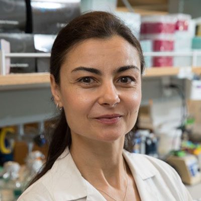
Roxana A. Radu
Associate Professor In-Residence
Retina Biochemistry and Disease Modeling
Dr. Radu's research investigates retinal degenerative diseases by exploring the retinal pigment epithelium (RPE), a crucial cell layer supporting photoreceptors. Using advanced genetic, biochemical, and cell biological approaches, including induced pluripotent stem cells and mouse models, the laboratory examines molecular mechanisms driving RPE-photoreceptor dysfunction, with the goal of developing potential therapeutic interventions.
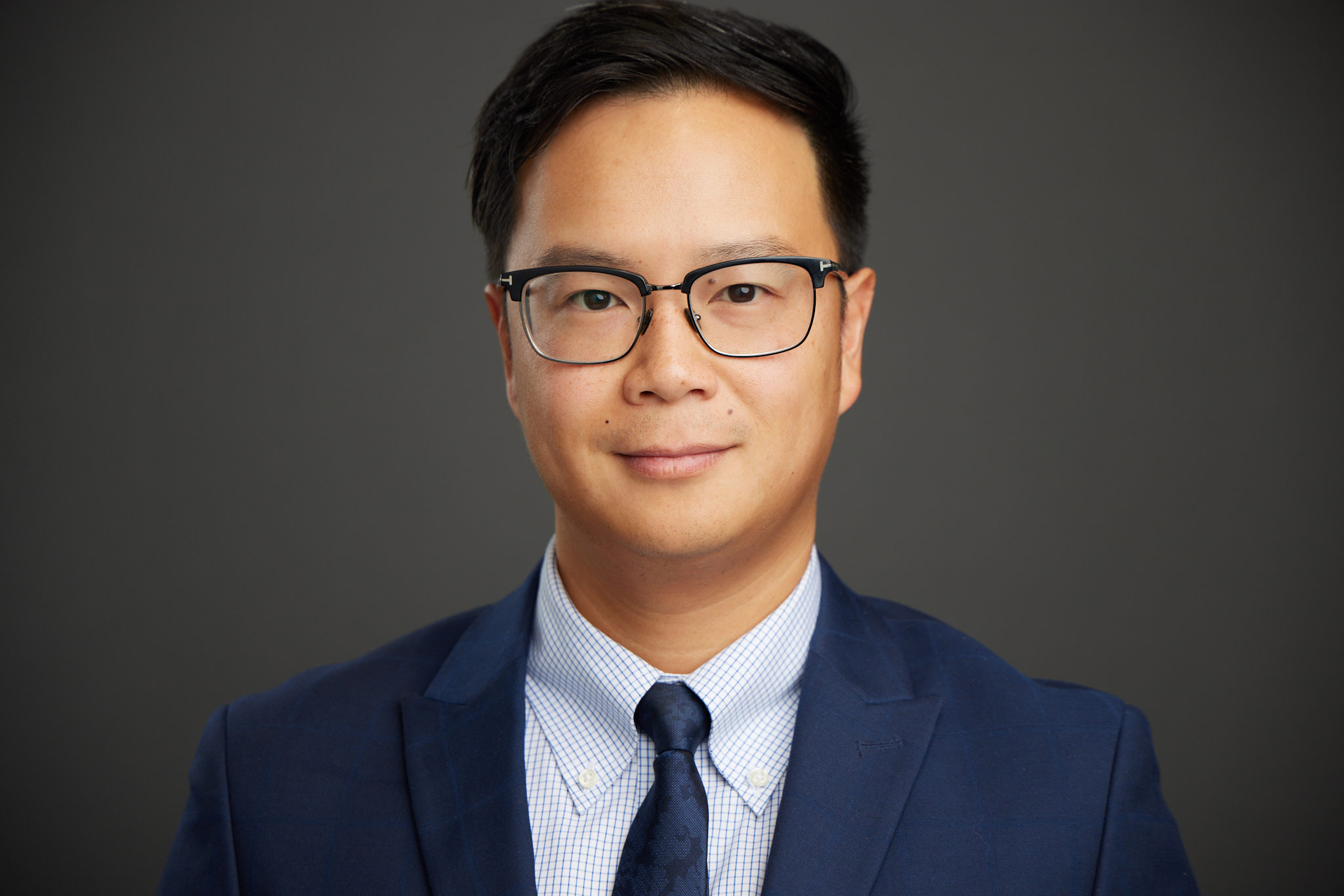
Edmund Tsui
Associate Professor
Uveitis and Ocular Inflammation
Dr. Tsui specializes in the study and treatment of ocular inflammatory diseases. His research focuses on advanced imaging analysis and biomarker discovery in uveitis. He also leads multiple clinical trials aimed at developing innovative therapies for uveitis and other inflammatory eye conditions.
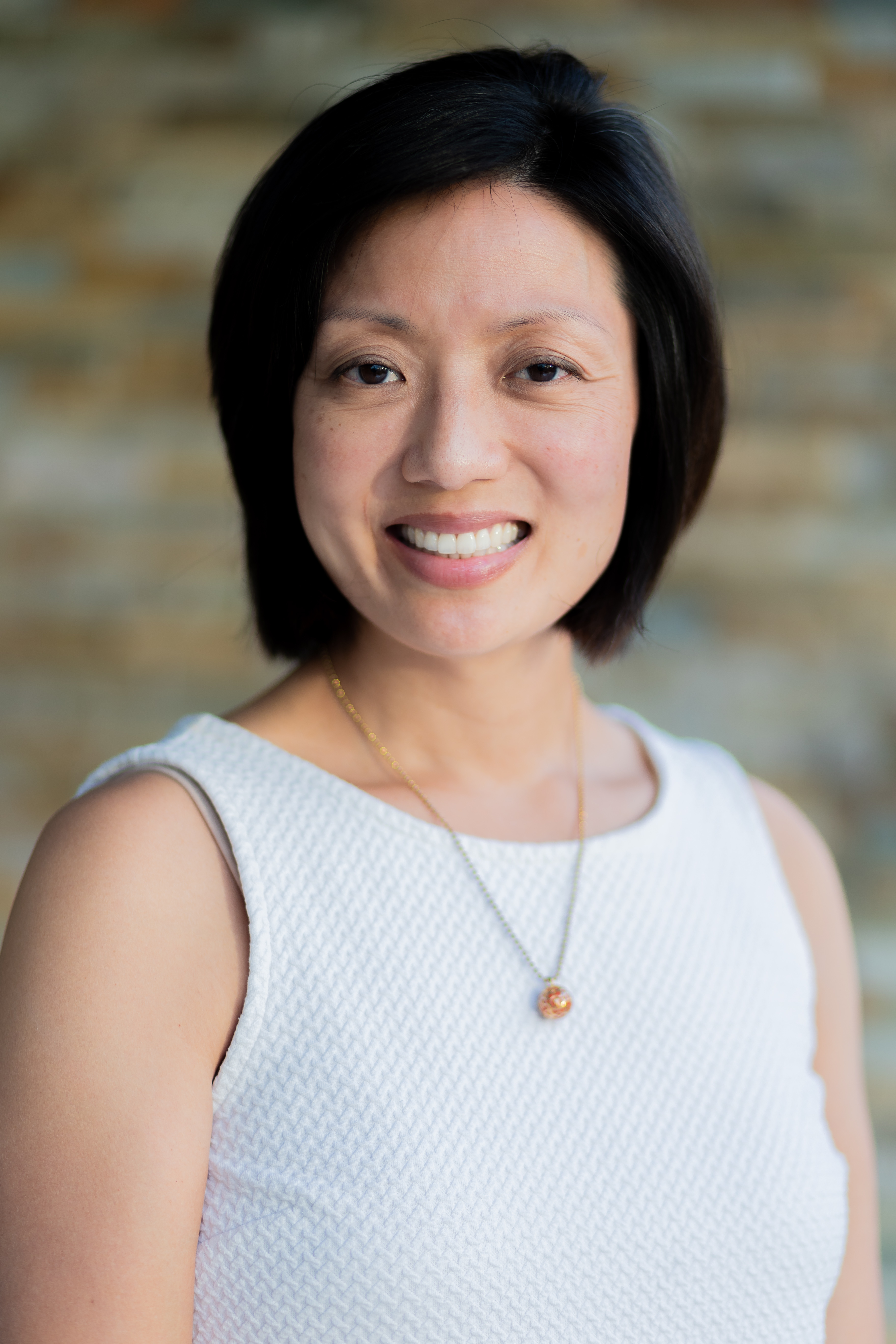
Irena Tsui
Associate Professor
Clinical Research in Retinal Disease
Dr. Tsui specializes in clinical research focused on retinal disease and oculomics. Her work combines clinical insights with optical coherence tomography angiography to evaluate systemic conditions, particularly in premature infants and pregnant women.
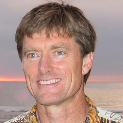
David Williams
Professor In-Residence
Retinal Cell Biology
The Williams laboratory studies dynamic cellular events in photoreceptor and RPE cells, with the aim of understanding basic cellular functions within these highly specialized cells, as well as the underlying processes in retinal degenerations.