David Williams Laboratory
Retinal Cell Biology

David S. Williams, Ph.D.
Professor
Karl Kirchgessner Foundation Chair in Vision Science
Departments of Ophthalmology and Neurobiology
Director, Microscopy and Image Analysis Core
Jules Stein Eye Institute
dswilliams@ucla.eduFaculty Profile
Laboratory WebsiteORCID Profile
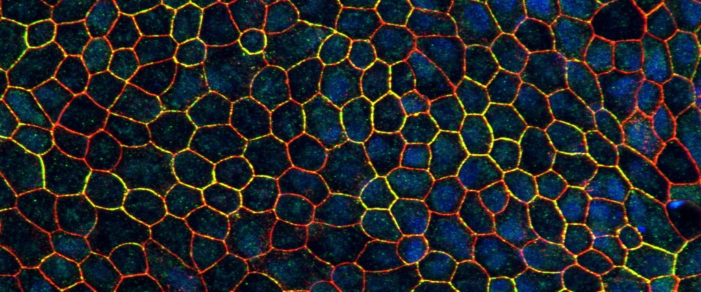
The Williams laboratory studies dynamic cellular processes in photoreceptor and RPE cells, with the aim of understanding basic cellular functions within these most highly specialized cells. Emphasis is on using advanced microscopy methods, including high-speed live-cell imaging and 3-D electron microscopy.
Current Research Areas:
- The delivery of opsin to the photoreceptor cilium and membrane shaping to form the disk membranes the photoreceptor outer segment.
- RPE cell biology, especially the motility and interactions of organelles, such as mitochondria, melanosomes, phagosomes and endolysosomes.
- Cell metabolism in relation to RPE cell differentiation.
- Retinal degeneration in Usher syndrome, involving the differentiation of patient-derived iPSCs into retinal cells, studies of MYO7A, and strategies for treatments via gene therapy.
- Mechanisms of other retinal degenerations, including choroideremia and macular degeneration.
By studying fundamental cellular processes that are essential for photoreceptor and RPE cell function, the lab's research is advancing general cell biological principles. It also provides an understanding of how defects in these processes lead to retinal degeneration. This knowledge is critical for developing treatments for inherited retinal diseases.
Recent Publications
Ultrastructural and Electrophysiological Determination of the Patency of Monkey Cone Outer Segment Membranes
Antonio E. Paniagua, Stefanie Volland, Gregory S. Bryman, Gabriel Luna, Andrew W. Chang, Michael Tri H. Do, Steven K. Fisher, David S. Williams
Journal of Neuroscience (2026)
View PublicationCollaborative role of two distinct cilium-specific cytoskeletal systems in driving Hedgehog-responsive transcription factor trafficking
David S. Williams, Radhika Subramanian, Pei-I Ku, Jamuna S Sreeja, Abhishek Chadha, Martin F Engelke
Science Advances (2025)
View PublicationMachine learning-based 3D segmentation of mitochondria in polarized epithelial cells
Nan W. Hultgren, Tianli Zhou, David S. Williams
Mitochondrion (2024)
View PublicationProductive infection of the retinal pigment epithelium by SARS-CoV-2: Initial effects and consideration of long-term consequences
Nan W Hultgren, Anton Petcherski, Simona Torriano, Ravikiran Komirisetty, Madhav Sharma, Tianli Zhou, Barry L Burgess, Jennifer Ngo, Corey Osto, Byourak Shabane, Orian S Shirihai, Theodoros Kelesidis, David S Williams
PNAS Nexus (2024)
View PublicationCones and cone pathways remain functional in advanced retinal degeneration
Erika M. Ellis, Antonio E. Paniagua, Miranda L. Scalabrino, Mishek Thapa, Jay Rathinavelu, Yuekan Jiao, David S. Williams, Greg D. Field, Gordon L. Fain, Alapakkam P. Sampath
Current Biology (2023)
View PublicationExpression of two major isoforms of MYO7A in the retina: Considerations for gene therapy of Usher syndrome type 1B
W. Blake Gilmore, Nan W. Hultgren, Abhishek Chadha, Sonia B. Barocio, Joyce Zhang, Oksana Kutsyr, Miguel Flores-Bellver, M. Valeria Canto-Soler, David S. Williams
Vision Research (2023)
View PublicationThree-Dimensional Ultrastructure of the Normal Rod Photoreceptor Synapse and Degenerative Changes Induced by Retinal Detachment
Gil Torten, Steven K. Fisher, Kenneth A. Linberg, Gabriel Luna, Guy Perkins, Mark H. Ellisman, David S. Williams
The Journal of Neuroscience (2023)
View PublicationSpatiotemporal Live-Cell Analysis of Photoreceptor Outer Segment Membrane Ingestion by the Retinal Pigment Epithelium Reveals Actin-Regulated Scission
Ankita Umapathy, Gil Torten, Antonio E. Paniagua, Julie Chung, Madeline Tomlinson, Caleb Lim, David S. Williams
The Journal of Neuroscience (2023)
View PublicationCell culture models to study retinal pigment epithelium-related pathogenesis in age-related macular degeneration
Kapil Bharti, Anneke I. den Hollander, Aparna Lakkaraju, Debasish Sinha, David S. Williams, Silvia C. Finnemann, Catherine Bowes-Rickman, Goldis Malek, Patricia A. D'Amore
Experimental Eye Research (2022)
View PublicationVitamin B3, nicotinamide, enhances mitochondrial metabolism to promote differentiation of the retinal pigment epithelium
Roni A. Hazim, Antonio E. Paniagua, Lisa Tang, Krista Yang, Kristen K.O. Kim, Linsey Stiles, Ajit S. Divakaruni, David S. Williams
Journal of Biological Chemistry (2022)
View Publication