Microscopy and Image Analysis
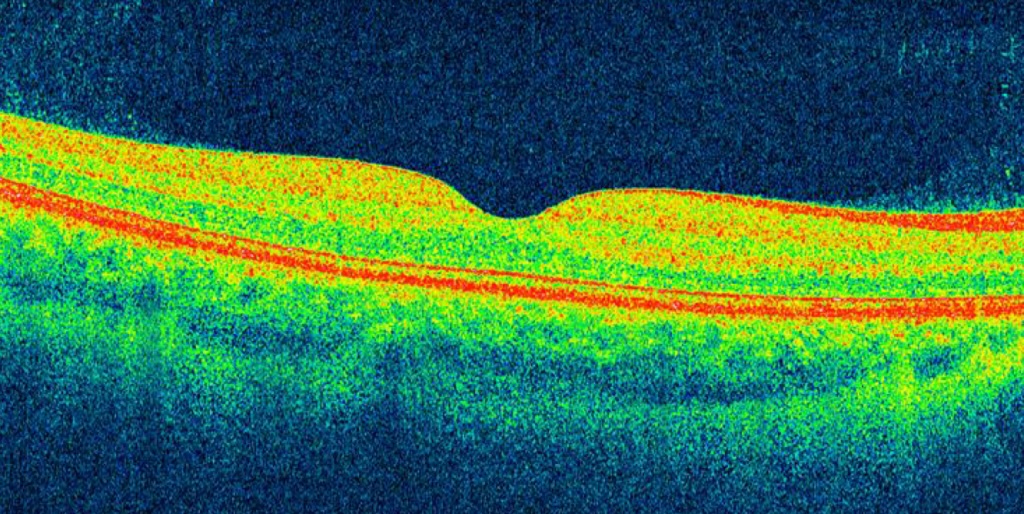
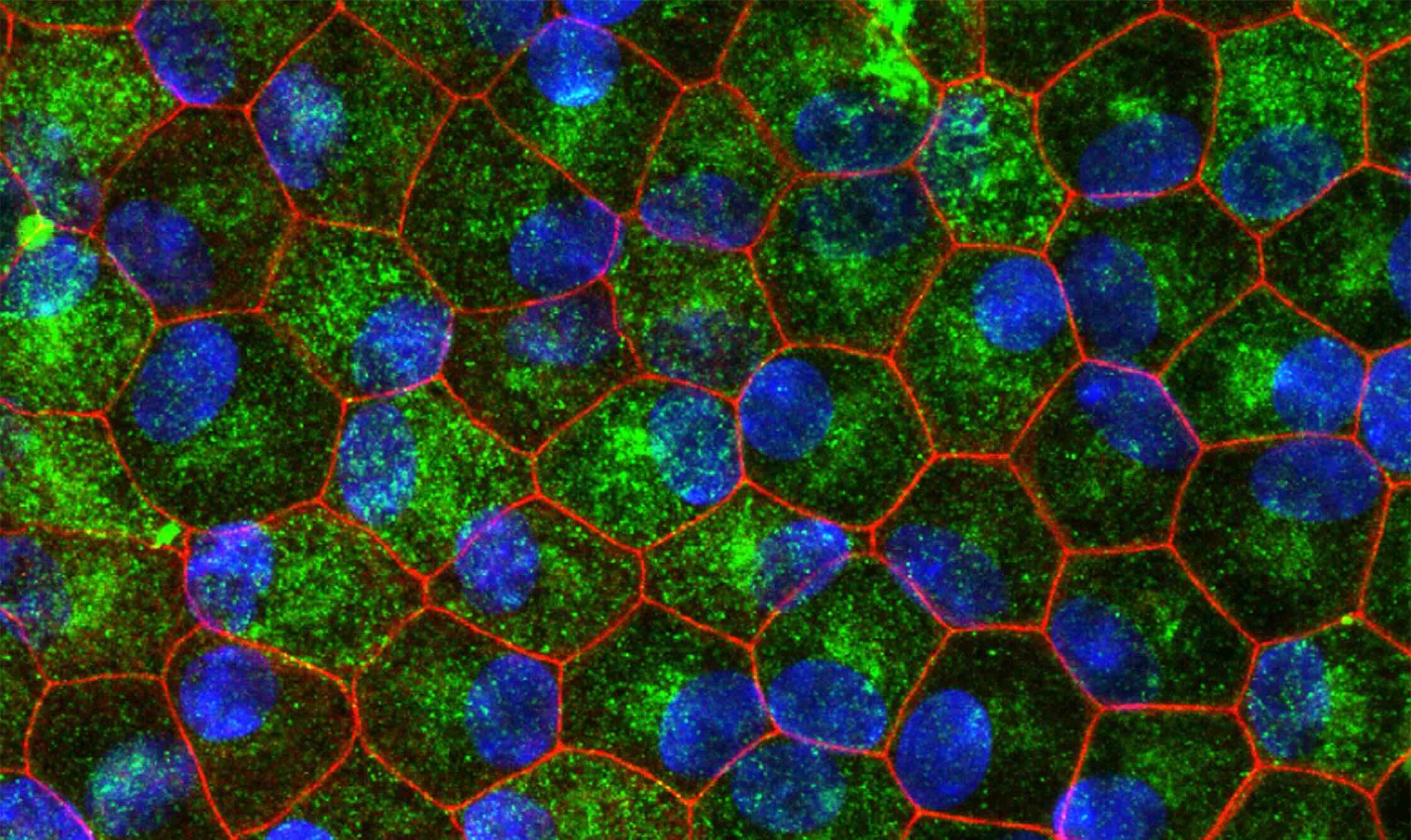
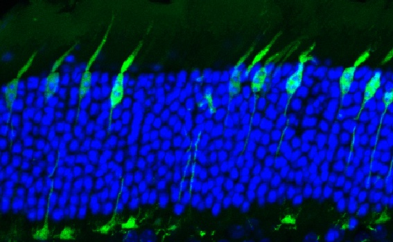
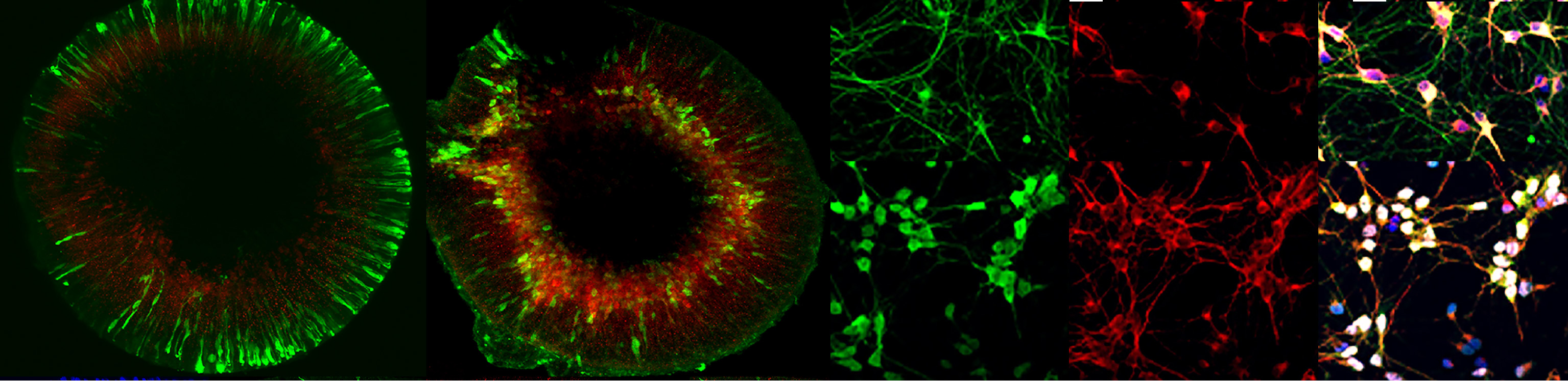
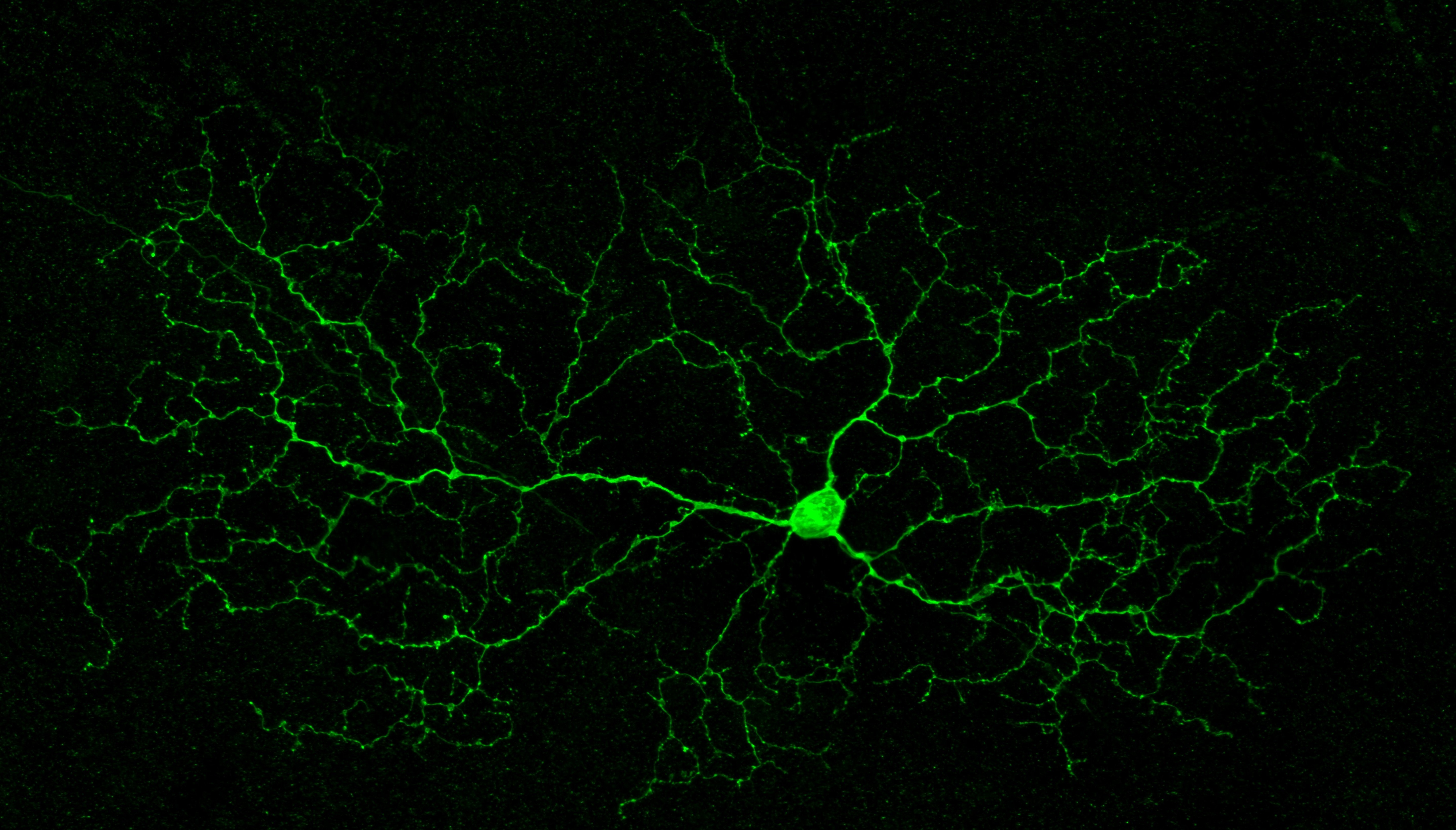
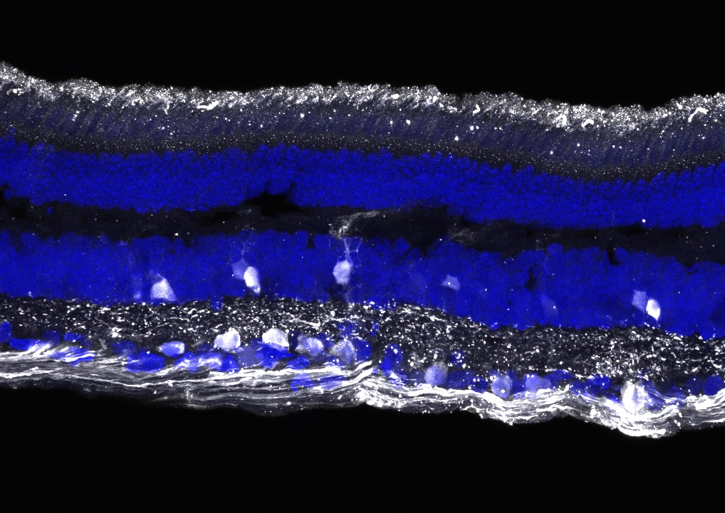
State-of-the-art microscopy for studies of retinal structure and function — from high-resolution confocal and super-resolution to electron microscopy and advanced 3D image analysis, including live-cell imaging.
- Director
- David Williams, Ph.D.
- Staff
- Yuekan Jiao, Ph.D. & Jane Coffman, Ph.D.
Key capabilities
- Wide-field and high-resolution microscopy
- Tissue and sample preparation
- Advanced image analysis, including 3D reconstruction