Joseph L. Demer Laboratory
Ocular Motility and Vision
Understanding eye movement control and strabismus

Joseph L. Demer, M.D., Ph.D.
Professor
Arthur L. Rosenbaum, MD, Chair in Pediatric Ophthalmology
Departments of Ophthalmology and Neurology
Director, Prototype Construction Core
Jules Stein Eye Institute
jld@jsei.ucla.eduFaculty Profile
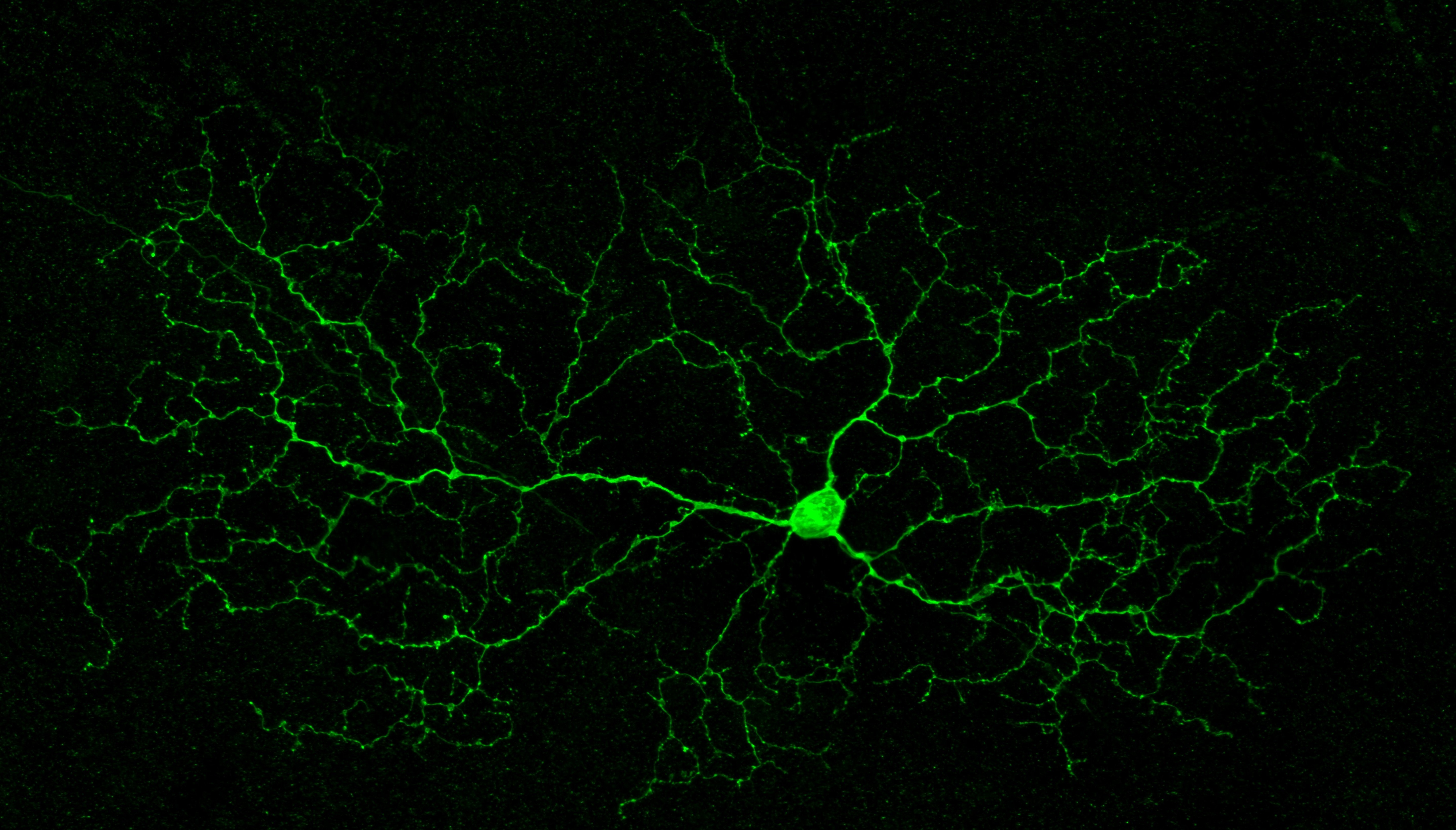
While the extraocular muscles obviously play complex roles in ocular rotation and alignment, extraocular muscle forces must mechanically balance against the eyeball, optic nerve, and a network of suspensory tissues within the orbit. The effects of eye rotation are concentrated at the optic disc and propagate from it into surrounding retina, choroid, and sclera. The optic nerve both contributes to muscle loading, and is in turn loaded by the muscles, potentially contributing to: 1) strabismus, the misalignment of the eyes often associated with double vision; 2) normal tension glaucoma associated with visual loss; and 3) pathologic myopia, the elongation and deformation of the eye causing nearsightedness. The Demer lab studies the dynamic ocular motility contribution to these disorders.
The Demer Lab studies mechanisms of strabismus by detailed clinical measurements of ocular motility in normal volunteers and patients with specific forms of strabismus such as superior oblique palsy and masquerading conditions. Specialized magnetic resonance imaging (MRI) technique quantitatively demonstrates the anatomy and function of the extraocular muscles and associated connective tissues in the eye sockets. Lumped parameter and finite element computational models are used to test hypotheses about mechanisms of strabismus, and to guide precision diagnosis and optimize surgical therapy for strabismus. Analysis of long-term results of standardized strabismus surgeries are used to develop and test quantitative, data-driven recommendations for use by surgeons worldwide. To date, these studies have fundamentally changed the understanding of the anatomy of human muscles and associated connective tissues, elucidating common but heretofore unrecognized causes of common strabismus.
The Demer lab performs MRI and optical imaging of the human eye during eye movements to measure the resulting deformations of the optic nerve, optic disc, retina, and choroid. Tissue deformations determined from the imaging using machine learning are compared with finite element computational models based on measured biomechanical properties of post-mortem human ocular tissues. Resulting models are then correlated with clinical findings in patients with glaucoma and pathological myopia. These studies suggest that eye movements significantly to both common, blinding disorders, and may eventually improve their treatments.
Selected Publications
Eye movements and the intraorbital subarachnoid space in optic neuropathy
Demer JL, Clark RA, Suh SY, Giaconi JA, Nouri-Mahdavi K, Law SK, Bonelli L, Arnold AC, Quiros P, Coleman A, Caprioli J
Invest. Ophthalmol. Vis. Sci (2025) • in press
View PublicationPostmortem digital image correlation and finite element modeling demonstrate posterior scleral deformations during optic nerve adduction tethering
Lim S, Kim K, Jafari S, Park J, Garcia SS, Demer JL
Bioengineering (2024)
View PublicationOptical coherence tomography angiography demonstrates strain and volume effects on optic disc and peripapillary vasculature caused by horizontal duction
Lim S, Tran A, Garcia SS, Demer JL
Cur. Eye Res (2023)
View PublicationScanning laser ophthalmoscopy demonstrates disc and peripapillary strain during horizontal eye rotation in adults
Park J, Moon S, Lim S, Demer JL
Am. J. Ophthalmol (2023) • 254: 114-127
View PublicationOptic nerve traction during adduction in open angle glaucoma with normal versus elevated intraocular pressure
Demer JL, Clark RA, Suh SY, Giaconi J, Nouri-Mahdavi K, Law SK, Bonelli L, Coleman AL, Caprioli J
Cur. Eye Res (2020) • 5(2): 199-210
View Publication